Metallurgical Abstracts on Light Metals and Alloys vol.57
Effect of Hydroxyapatite Coating Thickness on Inflammation and Osseointegration of Ti-29Nb-13Ta-4.6Zr (TNTZ) implants
Nuzul Ficky Nuswantoro*, Gunawarman**, Menkher Manjas***, Netti Suharti****, Dian Juliadmi*, Nila Kasuma*****, Yusril Yusuf******, Aminatun*******,
Yessie Widya Sari********, Mitsuo Niinomi********* and Toshikazu Akahori**********
* Research Center for Biomass and Bioproducts, National Research and Innovation Agency
** Department of Mechanical Engineering Department, Universitas Andalas
*** Department of Surgery, M. Djamil Hospital
**** Department of Microbiology, Faculty of Medicine, Universitas Andalas
***** Faculty of Dentistry, Universitas Andalas
****** Department of Physic, Universitas Gajah Mada
******* Department of Physics, Universitas Airlangga
******** Department of Physics, Institut Pertanian Bogor
********* Institute for Materials Research, Tohoku University
********** Department of Material Science and Engineering, Meijo University
[Published in Journal of Journal of Materials Research and Technology, Vol. 30, (2024), 6210-6217]
https://doi.org/10.1016/j.jmrt.2024.05.013
E-mail: akahori[at]meijo-u.ac.jp
Key Words: beta type titanium alloy, implant, hydroxyapatite, osseointegration
The study investigated the effect of hydroxyapatite (HA) coating thickness on the inflammation and osseointegration properties of Ti-29Nb-13Ta-4.6Zr (TNTZ) implants for orthopedic implant applications. TNTZ screws were used as based samples, and the HA coating was deposited using an electrophoretic deposition (EPD) method to achieve various coating thickness. The thin (50-70 µm), medium (70-90 µm), and thick (90-110 µm) coating thicknesses were obtained by applying different voltage and time exposure during the EPD process, while the uncoated screw was used as the control sample. The screws were implanted into the right tibia of male Rattus norvegicus Wistar rats for two weeks. After termination of the test animals, blood samples were collected to measure levels of tumor necrosis factor-α (TNF- α) using an ELISA method, and a removal torque test was performed on the implanted screws. Additionally, tibial bone tissue around the implant area was collected for histopathology analysis. The result of the study indicated that HA coating thickness has effects on TNF-α concentration, removal torque value, and new bone growth. The optimal thickness of the HA layer was found to be in the medium range (70-90 µm), resulting in low inflammation levels, relatively high osseointegration rates, and optimal new bone tissue growth. These finding were expected to be applicable in future orthopedic implant applications.
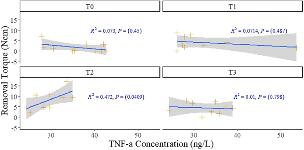
Correlation between TNF-α concentration and removal torque value in each coating thickness group.