Metallurgical Abstracts on Light Metals and Alloys vol.55
Microstructural Evolution in Magnesium after Hyper-Velocity Impact of Alumina Projectile
Naoki Fujita*,**, Tatsuya Nakatsuji**, Sunao Hasegawa***, Naoko Ikeo**, Eiichi Sato*** and Toshiji Mukai**
*Graduate Student, Graduate School of Engineering, Kobe University
**Graduate School of Engineering, Kobe University
***Institute of Space and Astronautical Science, Japan Aerospace Exploration Agency
[Published in Materials Transactions, Vol. 62 (2021), pp. 1401–1406]
https://doi.org/10.2320/matertrans.MT-L2021003
E-mail: Mukai[at]mech.kobe-u.ac.jp
Key Words: magnesium, hyper velocity impact, smoothed particle hydrodynamics method,
recrystallization, crack propagation
Magnesium specimens were impacted by a spherical alumina projectile at a velocity around 7 km/s under two environment temperatures of room temperature (~300 K) and low temperature (~173 K). To clarify deformation and fracture mechanisms, macro- and micro-structure were inspected by using micro-X ray computed tomography and scanning electron microscope (SEM) with electron back scattering diffraction (EBSD). In addition, simulation of the hyper-velocity impact was conducted using Smoothed Particle Hydrodynamics method to investigate the cumulative strain and temperature rise during the deformation. After a projectile impacted a target, a crater was formed on the target together with several cracks. In a closed portion below the crater formed at room temperature, fine grains and subgrains were observed by SEM/EBSD. From the calculation results, a temperature rise around 0.5 Tm (Tm; melting temperature of magnesium) and cumulated strain over 0.6 was suggested at 0.5 mm away from the edge of the crater. Therefore, the microstructure evolution was expected to be induced by the recrystallization and recovery due to the strain cumulated during the impact and the resultant temperature rise. On one hand, inspection of microstructure near the cracks revealed that microcracks were tended to propagate along grain boundary.
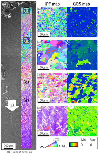
Microstructure of the magnesium target near the crater impacted at room temperature, (a) observed image with SEM, (b-e) IPF (left) and GOS (right) maps in the square portions indicated in (a).