Ti-15Zr-based alloys with Mo addition have been recently developed in order to achieve low Young’s modulus and non-cytotoxic chemical composition. Zirconium (Zr) presents similar chemical properties than titanium (Ti), which can improve mechanical strength, corrosion resistance and biocompatibility of the solid solution. Molybdenum (Mo) is a strong β-stabilizer, with tends to decrease Young’s modulus, still maintaining high mechanical strength. In our previous study, Zr composition has been set up with a binary Ti-Zr alloys and a Ti-Zr-Mo alloys have suited low Young’s modulus and adequate tribocorrosion resistance for biomedical applications. In this study, the tensile properties and cytocompatibility of the developed Ti-15Zr-xMo (x=0, 5, 10, and 15 wt%) alloys for biomedical applications were evaluated.
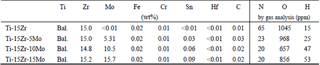
Ti-15Zr-based alloys were produced in an argon arc-melting furnace and molded in a centrifugal casting machine. The chemical composition and interstitial content of the samples were measured (Table 1). The mechanical properties were evaluated by tensile tests, Young’s modulus and Vickers microhardness measurements, being the results correlated with the corresponding phase composition and microstructure, which were analyzed by X-ray diffraction (XRD) and transmission electron microscopy (TEM) imaging. Cytocompatibility and cell proliferation were evaluated by MTT colorimetric assay.
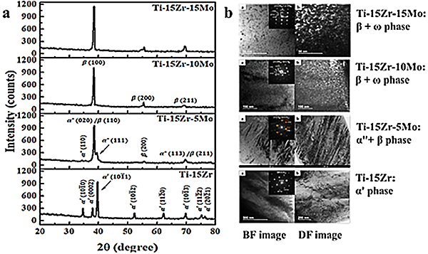
The phase composition of the samples was examined by XRD (Fig. 1a). The Ti-15Zr sample exhibited diffracted peaks from the hcp (martensitic α' phase) crystalline structure, whilst the Ti-15Zr-5Mo sample showed a biphasic composition composed of diffracted peaks from the orthorhombic (martensitic α" phase) and bcc (β phase) crystalline structures. The Ti-15Zr-10Mo and Ti-15Zr-15Mo samples presented only diffracted peaks from bcc crystalline structure, indicating that the amount of alloying elements was enough to retain the metastable β phase. In addition TEM images (Fig. 1b) confirmed XRD results. A general and inward view of the α' phase lamella in the Ti-15Zr sample was shown; and the α" phase needles dispersed along with the phase matrix was displayed in Ti-15Zr-5Mo sample. Regarding the Ti-15Zr-10Mo sample, images exhibited a general view of the intergranular region of the β phase with dispersion of some nanometric ω phase particles, with size less than 50 nm. Similarly, the Ti-15Zr-15Mo sample exhibited the same dispersion of the ω phase particle through the β phase matrix. It is worth to point out that its nanometric size turns difficult its identification by conventional XRD. In addition, this amount of ω phase on the samples can affect directly their mechanical properties once it is the hardest Ti phase.
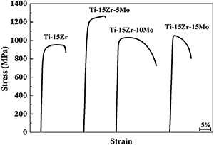
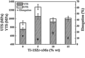
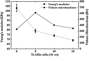
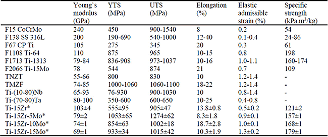
The representative stress-strain curve of the samples is compared in Fig. 2. Remarkable UTS values were observed in all samples. The Ti-15Zr-5Mo presented the highest UTS value, whilst Ti-15Zr-10Mo presented the largest elongation. The distinct tensile properties of the samples were a result of the different deformation modes of the Ti phases. The tensile properties of the samples are compared in Fig. 3. The Ti-15Zr-5Mo sample exhibited the highest UTS and YTS values, although its elongation was the lowest, possibly as a result of the α" phase precipitation on the β matrix. The Ti-15Zr-10Mo and Ti-15Zr-15Mo samples presented better combination of YTS and elongation values than the Ti-15Zr sample, which could be related to the distinct deformation mechanisms along the hcp and bcc crystalline structures. The Young’s modulus values (Fig. 4) exhibited a gradual reduction due to the β phase precipitation, which has the lowest value between the Ti phases. Because of the solution solid strengthening, all studied samples presented higher Vickers microhardness values than the CP Ti (Fig. 4). A comparison of the mechanical properties between the studied samples and some commercial metallic biomaterials are presented in Table 2.
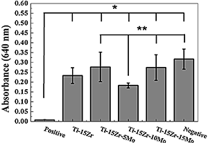
To evaluate the effects of particles and ions released into the culture medium by the samples, the indirect cell viability was obtained by the MTT assay after 72 h, being the extracts collected at 1g/mL from the medium after 48 h. In the indirect cytotoxicity test (Fig. 5) it can be noted that the number of viable cells in the presence of the extracts was high when compared to the positive control (growth medium with 1% phenol), which is also similar to the negative control (a normal growth medium). It can be concluded that the studied samples did not liberate any toxic component and neither affected the growth and cell viability during the studied time.
In this study, Ti-15Zr-based alloys with different amount of Mo were produced. The phase composition and microstructure were dependent on the alloying elements, being the Mo content of 5 wt% enough to precipitate the β phase. The Ti-15Zr-10Mo and Ti-15Zr-15Mo samples, composed by the β phase and small quantity of ω phase, presented similar mechanical strength, but distinct elongation and Young’s modulus values. All samples exhibited dimples-type structures along their fractured surfaces, which indicated a characteristic ductile behavior. Regarding a possible biomedical application, the Ti-15Zr-15Mo sample showed better mechanical biocompatibility than most commercial metallic materials, without any evidence of cytotoxicity on osteoblastic cells and proper wettability values. Therefore, the designed alloy could be suitable to mitigate the stress shielding effect, support the biomechanical loads of the body, and increase the lifespan of the implant without inducing cytotoxic effects.