Al-Al3Ti alloy containing Al3Ti particles with D022-type crystal structure is known as a grain refiner for as-cast aluminium and its alloys because the Al3Ti particles in the α-Al matrix play the role of good heterogeneous nucleation sites for α-Al. In a previous study, the Al-Al3Ti alloy was subjected to equal-channel angular pressing (ECAP) and its grain refining ability for as-cast Al was investigated. This Al-Al3Ti alloy contains platelet Al3Ti particles with a volume fraction of 11% in the matrix. When this alloy is deformed by ECAP, the number of Al3Ti particles increased due to their fragmentation. As a result, the Al-Al3Ti alloy deformed by ECAP had better grain refining ability compared with the undeformed Al-Al3Ti alloy. As well as ECAP, the cold rolling of the Al-Al3Ti alloy can enhance its grain refinement ability. Therefore, it is important to understand the fragmentation behavior of platelet Al3Ti.
Sato et al. have investigated fragmentation mechanism of the platelet Al3Ti particle in the Al-Al3Ti alloy by ECAP. As the result, it is found that the cracks in the platelet Al3Ti particles preferentially propagate along {112}Al3Ti when and Al3Ti particle in Al-Al3Ti alloy is fragmented by ECAP. However, because their results are obtained by microstructural analysis of fragmented Al3Ti particles, the fragmentation process of the platelet Al3Ti particles during deformation of the Al-Al3Ti alloy is still unclear. To clarify this process, it is necessary to perform the same area observation of the platelet Al3Ti particle during deformation for the Al-Al3Ti alloy. In this study, the fragmentation process of the platelet Al3Ti particle in Al-Al3Ti alloy during compression testing is investigated with the same area observation method.
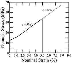
In order to investigate the microstructural change of a platelet Al3Ti particle in the Al-Al3Ti alloy during compression, the same area microstructural observation and compression testing were repeated. First, the initial microstructure of the Al-Al3Ti alloy before compression testing was observed by scanning electron microscopy (SEM) and electron backscatter diffraction (EBSD). After that, compression testing was performed to a nominal strain of 5% at a strain rate of 10-3/s. Subsequently, the microstructure of the platelet Al3Ti particle was observed using EBSD at the same position as the observation position for the initial microstructure. Furthermore, the compression test was carried out up to nominal strain of 8% using the same specimen, and then microstructural observation using EBSD is performed again at the same position.Figure 1 is a stress-strain curve obtained from compression testing of the Al-Al3Ti alloy. From the shape of this curve, the Al-Al3Ti alloy is greatly deformed by the compression test. Same-area observations of an Al3Ti particle were performed by stopping the compression test at nominal strains of ε = 5% and 8%.
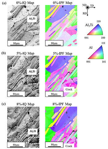
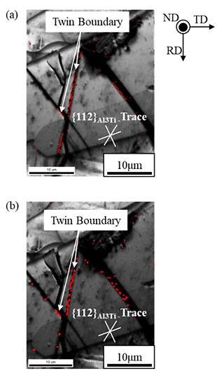
Figures 2 (a), (b) and (c) are image quality (IQ) map and inverse pole figure (IPF) maps of the Al-Al3Ti alloy specimens before and after compression testing up to nominal strains of 5% and 8%, respectively. At a nominal strain of ε = 5%, a crack is observed in the Al3Ti particle identified by the rectangle formed by dashed white lines. Furthermore, when the specimen was compressed up to ε = 8%, the Al3Ti particle was completely fragmented. Figures 3 (a) and (b) show highly magnified IQ maps of the specimen compressed up to nominal strains of ε = 5% and 8%, respectively. White lines in these figures are the traces of {112}Al3Ti. At ε = 5%, some boundaries and crack generation are formed on the platelet Al3Ti particle. These boundaries are parallel to the trace of {112}Al3Ti. Because of this, the boundary in Fig. 3 (a) is a deformation twin boundary. Furthermore, cracks propagated along the boundary parallel to the {112}Al3Ti trace, as shown in Fig. 3 (b). Therefore, it can be expected that deformation twinning occurred in the platelet Al3Ti particle during compression, and subsequently the crack propagated along the twin boundary with {112}Al3Ti.
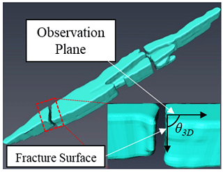
In order to investigate crystal plane orientation on fracture surface, 3-dimensitional (3D) reconstructed image of the fragmented Al3Ti particle was made by serial sectioning method. Figure 4 is 3D image of the fragmented Al3Ti particle. From crystallographic analysis and the shape of the reconstructed Al3Ti particle, it is found that the Al3Ti particles are preferentially fragmented along the {112}Al3Ti plane. Hence, it is concluded that the fragmentation of the platelet Al3Ti particles in Al-Al3Ti alloy preferentially occurs at twin boundary after deformation twinning.


